Archive
‘Cyborg’ spinal implant could help paralysed walk again
It might seem like science fiction but a new implant which attaches directly to the spine could help paralysed people walk again

The implant is so effective because it mimics the soft tissue around the spine so that the body does not reject its presence.
Paralysed patients have been given new hope of recovery after rats with severe spinal injuries walked again through a ‘groundbreaking’ new cyborg-style implant.
In technology which could have come straight out of a science fiction novel or Hollywood movie, French scientists have created a thin prosthetic ribbon, embedded with electrodes, which lies along the spinal cord and delivers electrical impulses and drugs.
The prosthetic, described by British experts as ‘quite remarkable’, is soft enough to bend with tissue surrounding the backbone to avoid discomfort.
Paralysed rats who were fitted with the implant were able to walk on their own again after just a few weeks of training.
Researchers at the Ecole Polytechnique Fédérale de Lausanne are hoping to move to clinical trials in humans soon. They believe that a device could last 10 years in humans before needing to be replaced.
The implant, called ‘e-Dura’, is so effective because it mimics the soft tissue around the spine – known as the dura mater – so that the body does not reject its presence.
“Our e-Dura implant can remain for a long period of time on the spinal cord or cortex,” said Professor Stéphanie Lacour.
“This opens up new therapeutic possibilities for patients suffering from neurological trauma or disorders, particularly individuals who have become paralyzed following spinal cord injury.”
Previous experiments had shown that chemicals and electrodes implanted in the spine could take on the role of the brain and stimulate nerves, causing the rats’ legs to move involuntarily when they were placed on a treadmill.
But this is the first study to show a simple gadget can help rats walk again and be tolerated by the body.
Scientists have struggled to find a device which will sit next to the spine or brain because both are surrounded by a protective envelope of tissue which the hard surface of implants can rub against, causing inflammation and scar tissue

The electronic ribbon is placed directly onto the spinal cord.
However the new gadget is flexible and stretchy enough that it can be placed directly onto the spinal cord. It closely imitates the mechanical properties of living tissue, and can simultaneously deliver electric impulses and drugs which activate cells.
The implant is made of silicon and covered with gold electric conducting tracks that can be pulled and stretched. The electrodes are made of silicon and platinum microbeads which can also bend in any direction without breaking.
Writing in the journal Science, where the results were published, science writer Robert Service said: “Soft flexible nerves connected to unyielding silicon and metal – the combination has spawned many a Hollywood cyborg.
“The implants Lacour’s team created still have to be wired to the outside world to operate, but she and her colleagues are designing wireless versions of the technology. Watch out, Hollywood, reality is catching up.”
The research was praised by British scientists.
“The work described here is a groundbreaking achievement of technology, which could open a door to a new era in treatment of neuronal damage,” said Dr Duško Ilić, Reader in Stem Cell Science at King’s College London.
“Until now, the most advanced prostheses in intimate contact with the spinal cord caused quite substantial damage to tissue in just one week due to their stiffness.
“There is still a long way to go before we may see any practical use of such neuroprostheses in humans. But it may be that it is something that could potentially be developed for use in humans in the foreseeable future.”
Prof John Hunt, Head of Unit of Clinical Engineering, University of Liverpool, added: “This study in rats is an interesting one and it could have the potential to be quite promising in terms of being applicable to people with spinal injuries.”
The implant has been primarily tested in cases of spinal cord injury in paralyzed rats but researchers believe it could eventually be used in epilepsy, Parkinson’s disease and pain management.
The scientists are planning to move towards clinical trials in humans within the next few years.
Additional Link:
NCBI – ‘Bionic’ spinal implant helped paralysed rats walk.
The research was published in the journal Science.
The above story is reprinted from materials provided by The Telegraph.
Striking the Cord: Optical Control of Motor Functions
Grad student Chi Lu and colleagues demonstrate a highly flexible polymer probe for triggering spinal-cord neurons with light and simultaneously recording their activity.
MIT researchers have demonstrated a highly flexible neural probe made entirely of polymers that can both optically stimulate and record neural activity in a mouse spinal cord — a step toward developing prosthetic devices that can restore functionality to damaged nerves.
“Our goal was to create a tool that would enable neuroscientists and physicians to investigate spinal-cord function on both cellular and systems levels with minimal impact on the tissue integrity,” notes Polina Anikeeva, the AMAX Assistant Professor in Materials Science and Engineering and a senior author of the paper published Nov. 7 in Advanced Functional Materials.
Department of Materials Science and Engineering graduate student Chi (Alice) Lu, who designed and implanted the probe, is the lead author of the study. Co-authors include Ulrich Froriep of the Simons Center for the Social Brain; Ryan Koppes of the Research Laboratory of Electronics; Andres Canales and Jennifer Selvidge of the Department of Materials Science and Engineering; and Vittorio Caggiano and Emilio Bizzi of the McGovern Institute for Brain Research. Professor Yoel Fink provided access to the fiber-drawing tower.
Experimental results
Although optogenetics, a method that makes mammalian nerve cells sensitive to light via genetic modification, has been applied extensively in investigation of brain function over the past decade, spinal-cord research has lagged. Earlier this year Caggiano and Bizzi have demonstrated inhibition of motor functions using optogenetics, and now the collaboration between the two groups yielded a device suitable for spinal optical excitation of muscle activity, while giving the researchers an electrical readout.
“Working in a spinal cord is significantly more difficult than in the brain because it experiences more movements. The radius of the mouse spinal cord is about 1 millimeter, and it is very soft, so it took some time to figure out how to design a device that would perform the stimulation and recording without damaging that tissue,” Lu explains.

Polymer fibers designed to mimic fibrous nerve geometry allow for simultaneous optical stimulation and neural recording in the spinal cord. This image illustrates the flexibility of the bifunctional neural probes and their ability to accommodate deformation due to the movement of vertebrae. Credit Chi (Alice) Lu and Polina Anikeeva.
The fiber was drawn from a template nearly 1.5 inches thick to its final diameter comparable to that of a human hair. It is flexible enough to be tied in a knot. The probe consists of a transparent polycarbonate optical core; parallel conductive polyethylene electrodes for recording neuronal electrical activity; and cyclic olefin copolymer acting both as electrical insulation and optical cladding. The flexible probe maintains its optical and electrical functions when bent by up to 270 degrees at very small radii of curvature (e.g. 500 µm), albeit with somewhat diminished light-carrying capacity at those conditions. The device still performed well after repeated bending and straightening, holding up under stresses expected from normal body movements, the report shows. MIT has filed a patent on the device platform.
The researchers conducted experiments with their neural probe in genetically-altered mice that express the light-sensitive protein channelrhodopsin 2 (ChR2) labeled with yellow fluorescent protein. The ChR2 makes neurons in the mice respond to blue light. These mice, developed by Professor Guoping Feng and colleagues at the McGovern Institute for Brain Research, provide a convenient model system for optoelectronic neural prosthetics. “When pulses of blue light are delivered to the spinal cord, we can directly observe neuronal response by getting an electrical recording,” explains Lu, who entered the third year of her doctoral program this fall.
“Laser pulses … delivered through the [polycarbonate] core of the fiber probe robustly evoked neural activity in the spinal cord, as recorded with the … electrodes integrated within the same device,” the researchers report.
The fiber was inserted into the proximal lumbar section of the spinal cord in mice, and light delivered through it triggered activity in one of the calf muscles, the gastrocnemius muscle. The results in the optically-sensitive mice were validated by comparison with results in wild type mice, which showed no response to the optical trigger. A toe pinch showed the device could still record mechanically stimulated neuronal activity in the wild-type mice. The researchers monitored muscle activity through electromyographical (EMG) recording, while the conductive polyethylene electrodes in the new device recorded neuronal activity in the spinal cord.
The MIT researchers’ combination in a single system of both recording activity from neurons and stimulating neurons with light is new, says Ravi V. Bellamkonda, the Wallace H. Coulter Professor and Department Chair of Biomedical Engineering at Georgia Institute of Technology and the Emory School of Medicine. “In principle, one would like to use ‘closed-loop’ systems, i.e., you detect a neurological event — like the brain wanting to move a limb — and then stimulate to affect that function when the natural link between them is severed due to an injury like spinal cord damage,” he explains.
“This is excellent engineering combining electrical and optical engineering for an important biological application — modulation of neural function in a closed-loop way. I am eager to see this technology being used in a biologically significant ways in the future,” Bellamkonda says.
The work was funded in part by grants from the National Science Foundation through the Center for Sensorimotor Neural Engineering and Center for Materials Science and Engineering; the McGovern Institute for Brain Research Neurotechnology Program; and the Simons Foundation.
Source: MIT press release
Image Source: The image is credited to the Chi (Alice) Lu and Polina Anikeeva and is adapted from the MIT press release
Original Research: Abstract for “Polymer Fiber Probes Enable Optical Control of Spinal Cord and Muscle Function In Vivo” by Chi Lu, Ulrich P. Froriep, Ryan A. Koppes, Andres Canales, Vittorio Caggiano, Jennifer Selvidge, Emilio Bizzi and Polina Anikeeva in Advanced Functional Materials. Published online August 26 2014 doi:10.1002/adfm.201401266
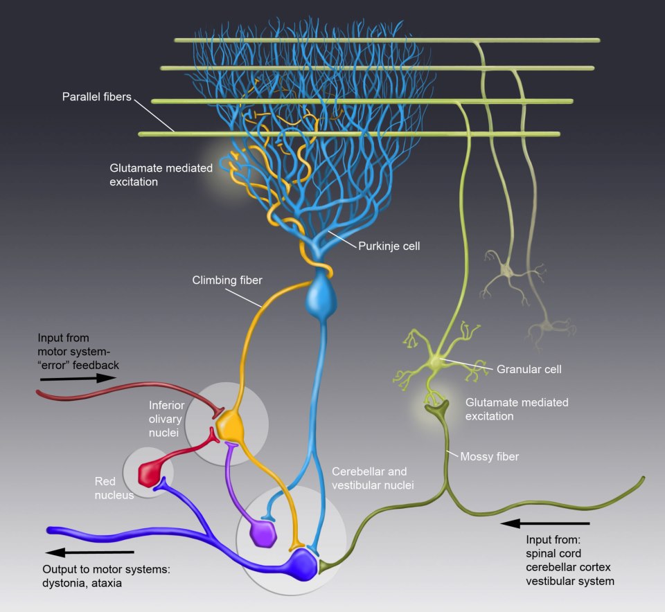
Neuronal Connections in the Cerebellum in Short
Control of movement is largely determined by incoming (afferent) and outgoing (efferent) neural impulses in the cerebellum.
Motor information input travels from the spinal cord, cerebral cortex and vestibular system via mossy fibers.
Feedback regarding movements returns to the cerebellum via the inferior olivary nucleus in the medulla oblongata. This feedback loop allows the brain to coordinate movement.
All outgoing neural impulses from the cerebellum travel via the deep cerebellar and vestibular nuclei. Proper functioning of the neuronal pathway between mossy fibers, granular cells, parallel fibers, climbing fibers and Purkinje cells are thought to be essential for coordinated muscular movement. Glutamate is a neurotransmitter in the excitatory synapses between climbing fibers and Purkinje cells as well as between granular cells and mossy fibers. Disruptions in this system are thought to be involved in a variety of movement disorders.
 (click on the picture to view full size)
The Reticular Formation, Limbic System and Basal Ganglia
The Reticular Formation
It’s a ‘diffuse net’ which is formed by nerve cells and fibers. It extends from the neuroaxis spinal cord through medulla, pons, midbrain, subthalamus, hypothalamus and thalamus (spinal cord is relayed superiorly to the cerebral cortex).
Many afferent and efferent pathways project in and out of the RF from most parts of the CNS. The main pathways through the RF is poorly defined and difficult to trace using silver stains. Reticular formation can be divided into three columns : median, medial and lateral columns.
Functions of the Reticular formation
1. Control of skeletal muscles:
- RF modulates muscle tone and reflex activities (via reticulospinal and reticulo bulbar tracts). It is important in controlling muscles of facial expression when associated with emotions.
2. Control somatic and visceral sensation (influence can be excitatory or inhibitory)
3. Control of autonomic nervous system
4. Control of endocrine nervous system (hypothalamus and the pituitary)
5. Influence on the biological clock (rhythm)
6. The reticular activating system (arousal and level of consciousness are controlled by the RF)
Clinical note
When a person smiles for a joke, the motor control is provided by the RF on both side of the brain. The fibers from RF is separated from corticobulbar pathway (supply for facial muscles). If a patient suffers a stroke that involves corticobulbar fibers, he or she has facial paralysis on the lower part of the face, but is still able to smile symmetrically.
The Limbic System
| Limbic structures | Functions of the limbic system |
|
1. Influence the emotional behavior:a. Reaction to fear and angerb. Emotions associated with sexual behavior
2. Hippocampus is involved in converting short term memory to long term memory (If the hippocampus is damaged, patient is unable to store long term memory – Anterograde amnesia) |
The Basal Ganglia and their connections


Connections of the Basal Ganglia

Yellow arrow : Pallidofugal fibers
Caudate nucleus and the Putamen: main sites of receiving inputs
Globus pallidus: main site from which output leaves
Afferent and Efferent fibers
| Connections of the caudate nucleus and Putamen | Connections of the Globus pallidus | ||
| Afferent | Efferent | Afferent | Efferent |
| CS: CorticostriateTS: Thalamostriate
NS: Nigrostriate BS: Brainstem striatal fibers |
SP: Striatopallidalfibers
SN: Striatonigral fibers |
SP: Striatopallidalfibers | Pallidofugalfibers |
Functions of the Basal Nuclei
Basal Nuclei controls muscular movements by influencing the cerebral cortex (it doesn’t have direct control through descending pathways to the brainstem and spinal cord). It helps to prepare for the movements (enables the trunk and limbs to be placed in appropriate positions before discrete movements of the hands and feet).
Functional connections of the Basal Nuclei and how they influence muscle activities

REFERENCES:
1. Ben Greenstein, Ph.D, Adam Greenstein, BSc (Hons) Mb, ChB Color Atlas of Neuroscience
2. Allan Siegel Ph.D, Hreday N. Sapru Ph.D Essential Neuroscience, 1st Edition
3. Stanley Jacobson, Elliot M. Marcus Neuroanatomy for the Neuroscientist
4. Patrick f. Chinnery Neuroscience for Neurologists
5. Dale Purves Neuroscience, 3rd Edition
6. Suzan Standring Gray’s Anatomy
7. Keith L. Moore, Arthur F. Dalley, Anne M. R. Agur Clinically Oriented Anatomy
8. Frank H. Netter Atlas of Human Anatomy
9. Walter J. Hendelman, M.D., C.M. Atlas of Functional Neuroanatomy
10. Mark F. Bear, Barry W. Connors, Michael A. Paradiso Neuroscience Exploring the Brain
11. Dale Purves et al. Principles of Cognitive Neuroscience
12. Eric R. Kandel et al. Principles of Neural Science
Descending Tracts
Descending tracts have three neurons:
1. 1st order neurons (UMN): cell bodies are in the cerebral cortex and other supra spinal areas
2. 2nd order neurons: short and situated in the anterior grey column of the spinal cord
3. 3rd order neuron (LMN): situated in the anterior grey column and innervate the skeletal muscles through anterior roots of the spinal nerves
Corticospinal tract: rapid, skilled and voluntary movements
1st order neuron
Axons arise from the pyramidal cells of the cerebral cortex (situated in the 5th layer), 2/3 from the pre central gyrus and 1/3 from the post central gyrus:
1. 1/3 of fibers arise from the 1stry motor cortex (Area 4)
2. 1/3 of fibers arise from the 2ndry motor cortex (Area 6)
3. 1/3 of fibers arise from the parietal lobe
(Area 1, 2 and 3).
Descending fibers converge in the corona radiata and pass though the posterior limb of the internal capsule; organization of fibers within the internal capsule:
1. close to genu (medial): concerned with the cervical parts of the body
2. away from the genu (lateral): concerned with the lower extremity.
The tract then passes through the middle 3/5 of the basis pedunculi of the midbrain; organization of fibers in the midbrain:
- medially: cervical parts of the body
- laterally: lower limbs.
When the tract enters the pons, it’s broken into many bundles by the transverse pontocerebellar fibers. In the medulla oblongata, the bundles group together to form the pyramids. At the junction of the MO and the spinal cord, most fibers cross the midline at the decussation of the pyramids and enter the lateral white column of the spinal cord to form the lateral corticospinal tract (LCST). LCST descends length of the spinal cord and terminates in the anterior grey column of all the spinal segments.
The fibers which didn’t cross, descend in the anterior white column of the spinal cord as the anterior corticospinal tract (ACST). Fibers of the ACST eventually cross and terminate in the anterior grey column of the spinal cord segments in the cervical and upper thoracic regions.
2nd order neuron:
It’s an internuncial neuron.
3rd order neuron:
It’s a alpha or gamma motor neuron.
To read more click on this link to the full article: Descending Tracts
Ascending Tracts
Introduction
They are located in the white matter and conduct afferent information (may or may not reach consciousness). There are two types of information:
- Exteroceptive : originates from outside the body (pain, temperature and touch
- Proprioceptive : originates from inside the body (from muscles and joints)
Normally there are three neurons in an ascending pathway:
- 1st order neuron: cell body is in the posterior root ganglion
- 2nd order neuron: decussates (crosses to the opposite side) and ascends to a higher level of the CNS
- 3rd neuron: located in the thalamus and passes to a sensory region of the cortex
Pain and temperature pathway: lateral spinothalmic tract
1st order neuron
Peripheral process extends to skin or other tissues and ends as free nerve endings (receptors). Cell body is situated in the posterior root ganglion. Central process extends into the posterior grey column and synapses with the 2nd order neuron.
2nd order neuron
The axon crosses obliquely to the opposite side in the anterior grey and white commissures within one spinal segment of the cord. It ascends in the contralateral white column as the lateral spinothalamic tract (LSTT).
As the LSTT ascends through the spinal cord new fibers are added to the anteromedial aspect of the tract (sacral fibers are lateral and cervical fibers are medial). The fibers carrying pain are situated anterior to those conducting temperature.
As the LSTT ascends through the medulla oblongata, it’s joined by the anterior spinothalamic tract and the spinotectal tract and forms the spinal lemniscus. Spinal lemniscus ascends through the pons and the mid brain.
Fibers of the LSTT end by synapsing with the 3rd order neurons in the ventral posterolateral nucleus of the thalamus (here crude pain and temperature sensations are appreciated).
3rd order neuron
Axons pass through the posterior limb of the internal capsule and corona radiata to reach the somatosensory area in the post central gyrus of the cerebral cortex. From here information is transmitted to other regions of the cerebral cortex to be used by motor areas. The role of the cerebral cortex is interpreting the quality of the sensory information at the level of the consciousness.
Light (crude) touch and pressure pathway: anterior spinothalamic tract (ASTT)
1st order neuron
It is similar to the pain and temperature pathway.
2nd order neuron
The axon crosses obliquely to the opposite side in the anterior grey and white commissures within several spinal segments. It ascends in the contralateral white column as the anterior spinothalamic tract (ASTT). As the ASTT ascends through the spinal cord new fibers are added to the anteromedial aspect of the tract (sacral fibers are lateral and cervical fibers are medial).
As the ASTT ascends through the medulla oblongata, it’s joined by the lateral spinothalamic tract and the spinotectal tract and forms the spinal lemniscus. Spinal lemniscus ascends through the pons and the midbrain. Fibers of the ASTT end by synapsing with the 3rd order neurons in the ventral posterolateral nucleus of the thalamus (here crude awareness of touch and pressure sensations are appreciated).
3rd order neuron
Axons pass through the posterior limb of the internal capsule and corona radiata to reach the somatosensory area in the post central gyrus of the cerebral cortex. The sensations can be crudely localized. Very little discrimination is possible.
To read more click on this link to the full article: Ascending Tracts (pdf).
Blood supply of the spinal cord
Arterial supply : Supplied by 3 small arteries + feeder arteries

Segmental spinal arteries

ASA and PSA arteries are reinforced by segmental arteries, which enter the vertebral canal through the inter vertebral foramina. These arteries are branches of arteries outside the vertebral column (deep cervical, intercostal and lumbar arteries). Segmental arteries give rise to anterior and posterior radicular arteries.
Feeder arteries
Enter the vertebral arteries and anastomose with the ASA & PSA. The most important feeder artery is the Great anterior medullar artery (GAM) of Adamkiewicz. It arises from the aorta at lower thoracic or upper lumbar vertebral levels. This artery is unilateral. It lies in the left side of most people. It represents the major source of blood to the lower 2/3 of the spinal cord.
Veins of the spinal cord
Drain mainly into the veins of the brain and the venous sinuses via 6 tortuous longitudinal channels. Finally, they drain into the internal vertebral venous plexus.
Structure of the spinal cord (Grey matter & White matter)
Transverse section of the spinal cord:
Gray matter

White matter


Scribd
![]() is where my documents live!
is where my documents live!
